Submitted by James Klim DDS, CADStar host on 04/03/2020 - 4:51pm
Submitted by James Klim DDS, CADStar host on 03/18/2020 - 12:11pm
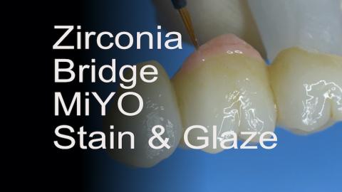
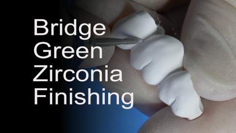
This video will demonstrate the aesthetic transformation of a posterior ZirCAD Multi bridge using the MiYO system by Jensen. The MIYO liquid ceramic system has transformed my ceramic and zirconia aesthetic clinical theater. It is a low fusing ceramic system and can be applied to all our CAD/CAM ceramic and zirconia systems. There are many assets to the MiYO system, but several of my favorites is that it is very easy to apply, multiple color combinations can be applied with one application, what is seen in the pre-fired state is what will be observed in the post-fired state, and the color characteristics appear to be embedded in the
Submitted by James Klim DDS, CADStar host on 03/18/2020 - 11:23am
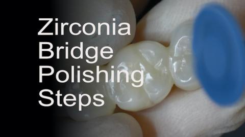
This is the third video in the posterior ZirCAD Multi bridge series. For the majority of my posterior bridges, I will polish only, unless the aesthetic blend requires more attention. This bridge will be placed in the upper left buccal corridor and is part of the secondary smile zone. MiYO by Jensen will be the stain and glaze product for the final characterizing phase. One thing that is nice about MiYO is that it can be applied to a highly polished surface. With this surface application characteristic, the zirconia bridge can be fully polished in preparation for the MiYO stain, glaze, and structure application.
Products used:
Submitted by James Klim DDS, CADStar host on 03/12/2020 - 10:52pm
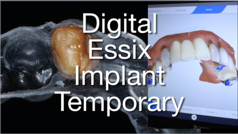
Primescan has made the full arch and full mouth scan a routine simple process in my clinical theater. This video will walk through the steps for processing an implant tooth Essix retainer. I currently have the Sprintray Pro and Asiga printers. They are both excellent printers and serve an important part in my daily practice. I will also share the products and method for tacking in the temporary tooth to the Essix retainer.
- Asiga or Sprintray Pro printer
- Ministar Forming Vacuum
- SR Connect (Ivoclar)
- Adhese Universal (Ivoclar)
- Flowable BulkFill (Ivoclar)
Submitted by James Klim DDS, CADStar host on 03/01/2020 - 10:43am
Part 2 for creating a posterior ZirCAD multi Bridge. Once the bridged is machined, what are the steps to effectively shape and finish the bridge before sintering? I follow a routine shaping workflow using the Meisinger JK04 Zirconia Lab Kit. Quite often, when using the multi zirconia options such as Katana or ZirCAD Multi, I will use selective infiltration to control value and enhance the multi-effect when the case demands a better blend.
- Meisinger JK04 Zirconia Lab Kit
- ZirCAD LT Coloring Liquids (Ivoclar)
Submitted by James Klim DDS, CADStar host on 02/16/2020 - 7:54pm
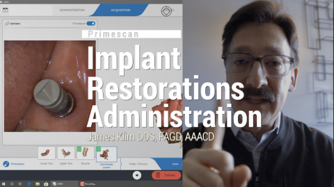
There are a few additional steps when setting up an implant restoration(s) in the administration screen. The first thing to check is affirming that the implant size and brand are in the current software system. Most mainline implant brands are. Before scheduling the patient, order the corresponding ScanPost. Scanning and fabricating an implant restoration is one of the most efficient and predictable applications in the CEREC software.
Submitted by James Klim DDS, CADStar host on 02/09/2020 - 8:55pm
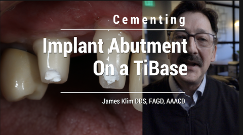
This video documents my current steps for cementing an implant abutment or one-piece implant restoration on the TiBase. The protocol that I have changed as of late is using Monobond Etch & Prime rather than hydrofluoric acid and silane. I have experienced great success with my TiBase created implant restorations. I have yet to see an e.max or zirconia abutment delaminated from the TiBase in 8 years.
Cementation Steps:
- Sandblast the adhesive surface on the TiBase
- MonoBond Plus metal priming on TiBase
- Fill access screw tube with plumbers tape
- MonoBond Etch & Prime internal adhesive
Submitted by James Klim DDS, CADStar host on 02/06/2020 - 9:30pm
Submitted by James Klim DDS, CADStar host on 01/21/2020 - 9:11pm
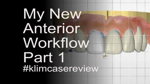
This video will highlight my anterior ceramic shaping workflow for multiple anterior restorations, particularly when crossing the midline. However, anterior restorative success is mostly about understanding the patient's treatment expectations and then working through the biomechanical diagnosis and preplanning with either a traditional or digital wax-up.
The predetermined wax-up prototype is used for preparation guidelines and reduction, transitional restorations when using a two appointment protocol, and a digital BioCopy scan for digital design guidance. When these preliminary and preparatory guidelines are delivered to the
Submitted by James Klim DDS, CADStar host on 12/16/2019 - 8:34pm
For posterior e.max restorations, spray glaze is one of the most effective ways to achieve an excellent luster finish. This video will walk through my spray glaze application technique for posterior e.max. For anterior teeth, I tend to apply brush-on glaze for more micro-surface texture control.
Make sure your whole team views this video, it is excellent for training and standardizing the e.max finishing workflow.

