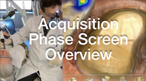
Number one reason to upgrade to 4.6 is the improved interpretation for rendered margin clarity. Also, there is improved responsiveness to acquiring ceramics, including e.max. The camera still struggles with shiny alloys. I have never found this a problem; I apply Optistray to decrease the reflective surfaces and acquire an incredible scan quality. This video will highlight the CEREC 4.6 Omnicam scanning qualities and my secret for staying on clinical schedule.
- Online Training
- New Content
Submitted by James Klim DDS, CADStar host on 11/11/2018 - 11:32am
Submitted by James Klim DDS, CADStar host on 11/09/2018 - 10:01am
Categories:
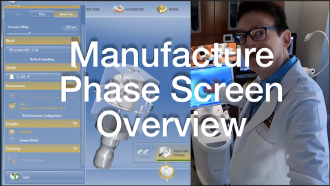
The Manufacture Palette graphic workflow has been upgraded. This is now where Occlusal Offset can be found and several other new screen links to easy access System Menu > Setting for milling bur change options and set default block size option.
Video Preview
Premium Member Full Video Viewing
Submitted by James Klim DDS, CADStar host on 11/08/2018 - 9:27pm
Categories:
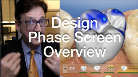
For "bread and butter" posterior applications, the virtual design process is seamless when there is enough occlusal reduction for the chosen material. This video will highlight my sequence for a smooth design phase screen flow.
Video Preview
Members Full Video
Submitted by James Klim DDS, CADStar host on 11/08/2018 - 1:03pm
Categories:
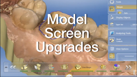
There are a few upgrades in the Model Phase Screen such as auto model axis and auto margin calculation. This video will review how to optimize these new features in the CEREC workflow.
Video Preview
Members Full Video
Submitted by James Klim DDS, CADStar host on 11/05/2018 - 7:37pm
Categories:

I am reaching more and more for Zirconia restorations for first and second molars when full coverage is indicated. With the current high strength zirconia, they tend to be opaque and often come into the room first! Since I have been color infiltrating in the green state, this simple efficient step has significantly improved the blend for the final finished restoration. This video will demonstrate my single zirconia restoration infiltration technique using ZirCAD LT Colouring Liquids.
Video Preview
Submitted by James Klim DDS, CADStar host on 10/26/2018 - 7:13am
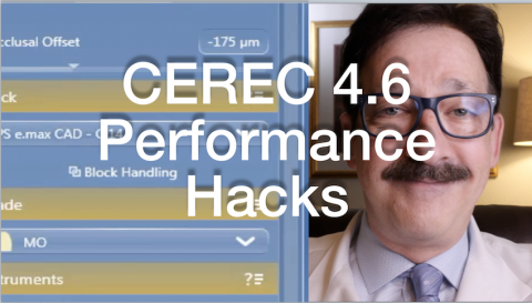
I recommend uploading CEREC 4.6 for just the scanning and margin clarity features. However, keep 4.52 around. There are historical trends for software glitches to be present with new software versions. This videos will share several hacks I use to get through some of the 4.6 glitches.
Video Preview
Video for Premium Members
Submitted by James Klim DDS, CADStar host on 10/21/2018 - 10:47pm
Categories:
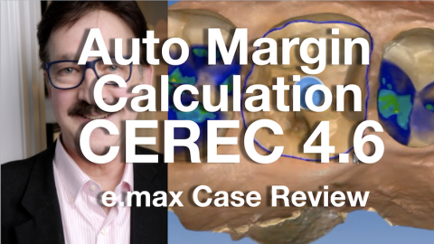
CEREC Case Review for auto margin calculation in the CEREC 4.6 Software, it draws the margin for you! When the margins are "high and dry", auto margin drawing is spot on. This case review will highlight the CEREC software for creating an e.max onlay restoration.
View Video Preview
View Member Full Video
Submitted by James Klim DDS, CADStar host on 10/19/2018 - 2:47pm
Categories:

When I need a high-strength zirconia restoration or bridge for the shadows of the mouth, I often reach for ZirCAD LT by Ivoclar. Avoiding visual tension is the secret for making high strength zirconia restorations work in the shadows. ZirCAD LT Colouring liquids are my secret. I using them on colored blocks to create a multi-effect and tone down the value in the cusp zones. This videos will show you how to set up the ZirCAD LT Colouring Liquid Kit.
Submitted by James Klim DDS, CADStar host on 10/16/2018 - 7:09am
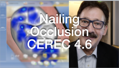
This video is about becoming a happy occlusal CAD/CAM clinician. There are five principles that I apply to achieve shimstock occlusion in CEREC 4.6. It is like clockwork. When you apply them, clinical theater life becomes much more pleasant.
Video Preview
Full Video for Members
Submitted by James Klim DDS, CADStar host on 10/07/2018 - 9:57pm
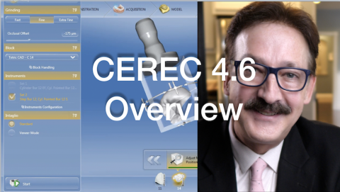
The CEREC 4.6 workflow is very similar to 4.52. The most significant upgrade from 4.52 is the camera scanning proficiency. The camera also picks up shiny surfaces such as ceramic and gold much better than in prior software versions. The model rendering is faster from the Acquisition screen and all the steps in the model screen are automated including setting Model Axis and Drawing Margins. I have found it is better to disable the auto margin calculation unless the margins are high and dry. For larger cases with multiple preparations, the software has a tendency to crash. The software performs much better when margin calculation is