- Online Training
- New Content
Submitted by James Klim DDS, CADStar host on 09/02/2014 - 9:47pm
Submitted by James Klim DDS, CADStar host on 08/27/2014 - 7:37pm
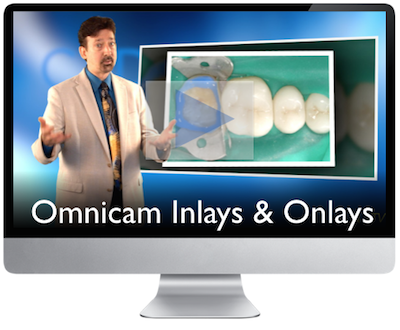
The Omnicam will now block out internal axial wall undercuts like Bluecam for inlay & onlays. For those that need this feature, Omnicam is now on board and will provide the block out options for more creative conservative preparations.
Submitted by James Klim DDS, CADStar host on 08/26/2014 - 9:37pm
Submitted by James Klim DDS, CADStar host on 08/24/2014 - 9:41pm
Submitted by James Klim DDS, CADStar host on 08/11/2014 - 5:41pm
Submitted by James Klim DDS, CADStar host on 07/27/2014 - 3:12pm
Submitted by James Klim DDS, CADStar host on 07/15/2014 - 3:04pm
Submitted by James Klim DDS, CADStar host on 06/04/2014 - 10:17pm
 Announcing a new video series posted in CEREC Integration Chapter One “Live in the CEREC Clinical Theater Omnicam”. Staying on schedule and achieving outstanding restorative results is depended on our clinical flow from the anesthetic approach, team delegation, digital imaging, software design, finishing, and cementation. It is often beneficial to step back and review the big picture of
Submitted by James Klim DDS, CADStar host on 06/02/2014 - 10:42pm
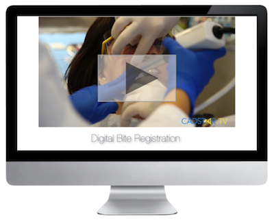 The digital bite registration is a very predictable record and will produce accurate occlusal contacts in the milled restoration. This tutorial will demonstrate how Dr. Klim effectively accomplishes this task. View video in Chapter 3
Submitted by James Klim DDS, CADStar host on 05/04/2014 - 9:44pm
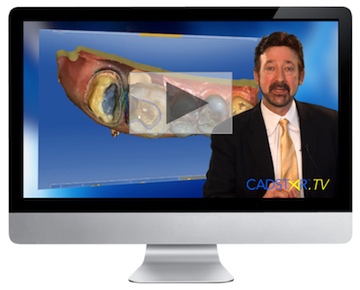 Omnicam will provide exceptional subgingival margin clarity. As of recent, I have been using gingival bur curettage for subgingival tissue retraction and then treating the soft tissue with Clear Hemostatic Gel to stop the bleeding. Within a short period of time, preparation and subgingival protocols are

